Institute of Engineering and Computational Mechanics
Biomechanical Investigation
Project Description
Figure 1: 3D model of the mandible.
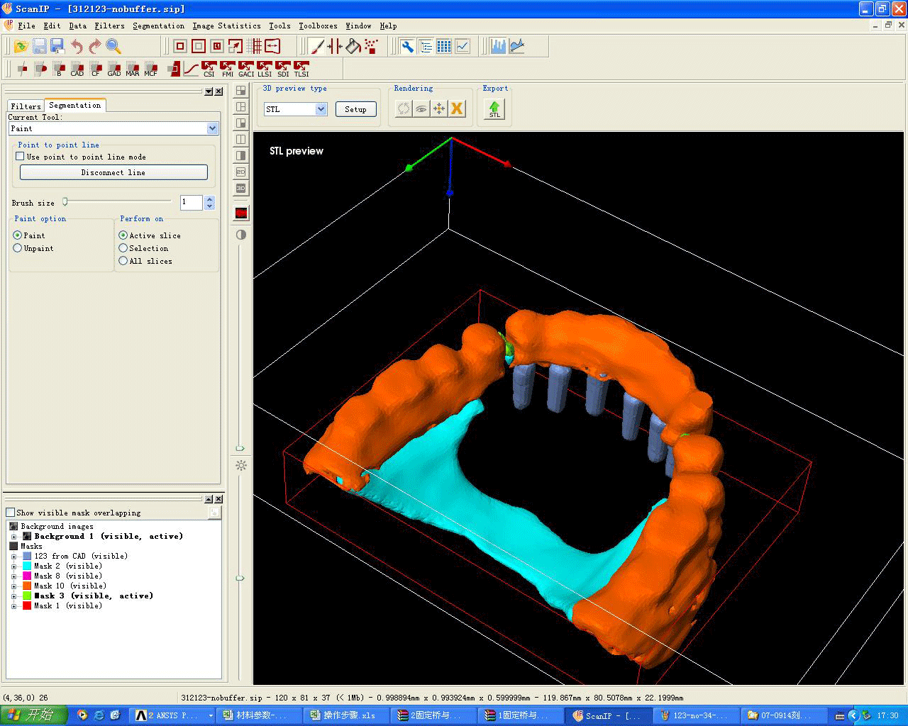
Figure 2: 3D model of edentulous maxilla restored with six anterior implants and resilient precision attachments placed distal to
canines.
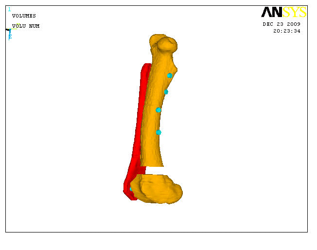
Figure 3: 3D model of distal femoral fractures treated with Less Invasive Stabilization System (LISS).
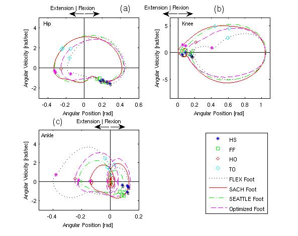
Figure 4: Phase Plane Portraits of Right Joints for Walking with Four Prostheses at Natural Speed.
The objective of project is the development of a method to construct a three-dimensional finite element model of the dentulous mandibular body of a normal person. A series of pictures were got by CT scanning. After extracting the cordinates of keypoints of some pictures, we constructed a platform of the three-dimensional finite element model of the dentulous mandibulare body. The three-dimensional model is constructed with lifelike shapes of dental cusps. Each part of this model can be easily removed.
Evaluation of Human Locomotion Characteristics for Different Prostheses and Walking Speeds.