The research project is to investigate the function and properties of incudo malleolaren-joint and their impact on the sound transmission of the middle ear. The IM-joint connecting the two middle ear malleus and incus and changed depending on the amplitude and frequency of the excitation to be transfer behavior. Under quasi-static displacements, as e.g. in the reconstruction or barometric pressure changes occur, shows a pronounced elastic flexibility of the joint. Under dynamic excitation frequency increases with increasing the influence of the viscosity of the synovial fluid in the joint, and thus improves the coupling. To characterize this behavior mechanical models are developed using which the relationships between the anatomical features of the IM-joint and its function should be examined systematically for the transmission of sound.
The project is funded by the DFG in the framework of the research program funded "transfer characteristics and function of incudo malleolaren-joint" and in collaboration with the University Hospital of Zurich (ORL).
Measurements
Dynamic excitation of the malleus-incus complex: the spatial velocity components can be determined with three laser Doppler vibrometers at several points. From this, the movement of the anvil can be reconstructed.
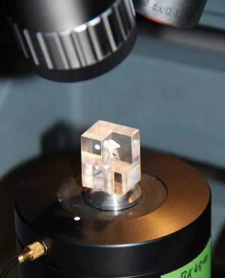
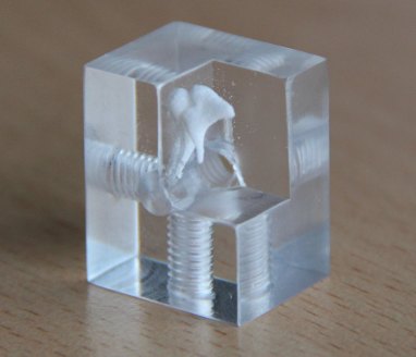
To characterize the mechanical properties of the IMG static and dynamic measurements are made on (isolated) malleus-incus complex. In static measurements forces are applied, and determines the shift between the ossicles. In the dynamic measurement of the malleus is excited by an electrodynamic vibration exciter (shaker), and determines the relative movement of the anvil. There are chosen different excitation parameters (direction, amplitude, frequency) and their influence investigated. From these measurements to the viscoelastic properties of the IM-joint can be derived. Is Furthermore, the frequency and amplitude response of the transmission behavior of the joint can be characterized.
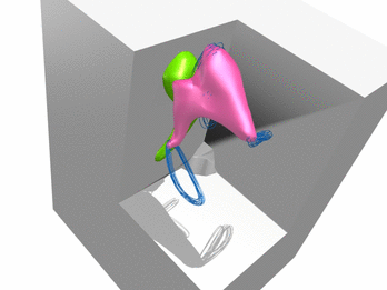
Visualization of the gap distance of the IM-joint of ZTB247 when a force is applied near the joint in lateral direction. The incus motion is guided by the geometric shape of the IM-joint. As the load is increased the gap gets smaller, with different region of the joint get into contact (highlighted in dark blue/grey).
Visualization of the gap distance of the IM-joint of ZTB247 when a force is applied at the long processus of incus in lateral direction. The incus motion is guided by the geometric shape of the IM-joint. As the load is increased the gap gets smaller, with different region of the joint get into contact (highlighted in dark blue/grey).
Visualization of the absolute value of the displacement of ZTB247 when applying a force at the long processus of incus in lateral direction. The grey color marks region with low values, the red color regions with larger values. The change of the instantaneous axis of rotation during the loading shows the influence of the joint geometry.
Comparison of the gap distance between measurement and simulation when a force is applied in lateral direction.
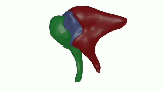
Motion of the middle ear ossicles when acoustically excited. The motion of the ossicles is reconstructed from 3D-LDV measurements of different points on the ossicles. The motion will occur at an excitation frequency of 500Hz with the motion being amplified.
Motion of the middle ear ossicles when acoustically excited. The motion of the ossicles is reconstructed from 3D-LDV measurements of different points on the ossicles. The motion will occur at an excitation frequency of 2000Hz with the motion being amplified.
Motion of the middle ear ossicles when acoustically excited. Beside the malleus and incus the temporal bone preparation is visible. The motion of the ossicles is reconstructed from 3D-LDV measurements of different points on the ossicles. The motion will occur at an excitation frequency of 850Hz with the motion being amplified.
Reconstruction of the orientation by markers attached to the temporal bone. The geometry is obtained from micro-ct scans of the temporal bone.
Modelling and Simulation
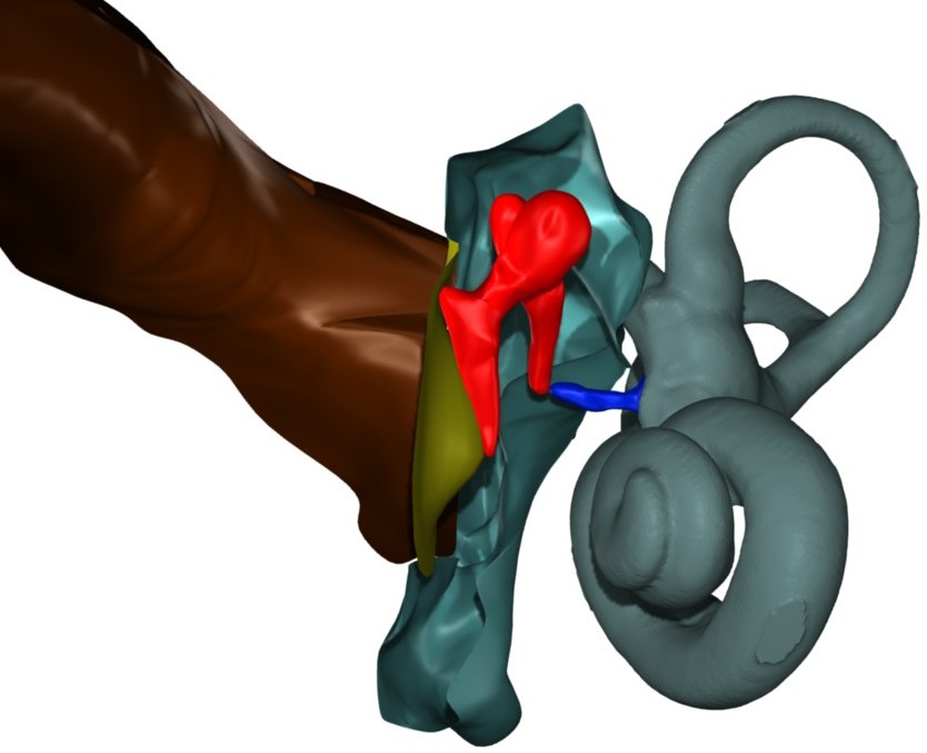
EMBS model of the human ear. Malleus and incus are colored in red.
With the aid of appropriate mathematical models replacement, the properties of the IM-joint will be described. These models can then be built into the embs model of human hearing.
The modelling of the elasticities is done by means of the Finite Element Method. The equations of motion of the coupled system are then derived using flexible multibody systems theory.